Today marks the 39th installment in a series of articles by HumanProgress.org titled Heroes of Progress. This bi-weekly column provides a short introduction to heroes who have made an extraordinary contribution to the well-being of humanity. You can find the 38th part of this series here.
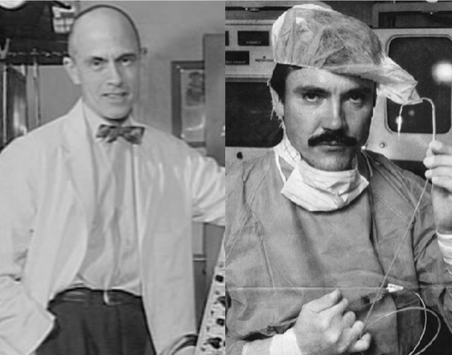
This week, our heroes are Charles Dotter and Andreas Gruentzig–two radiologists who pioneered angioplasty, which is a surgical procedure for widening narrow or blocked blood vessels. If left untreated, arterial atherosclerosis (i.e., plaque buildup in the veins) can cause severe medical problems, including coronary heart disease, heart attacks, strokes, etc. The World Economic Forum estimates that more than 15 million lives have been saved since Dotter’s initial discovery and Gruentzig’s subsequent improvement of angioplasty.
Charles Dotter was born June 14, 1920 in Boston, Massachusetts. Dotter attended a grammar school and high school in Freeport, Long Island. He was later described as a bright and inquisitive child. From an early age, Dotter was interested in mechanical objects and derived great satisfaction from dismantling and rebuilding machines. In 1941, Dotter was awarded a Bachelor of Arts degree from Duke University. In the same year, he enrolled in Cornell University’s medical school.
After medical school, Dotter completed an internship at the U.S. Naval Hospital in New York State, and later did his residency at New York University. At just 30 years old, Dotter was offered a job as a faculty member at Cornell Medical School and two years later he assumed the position of professor and chairman of the Department of Radiology at the University of Oregon. This new job meant Dotter became the youngest ever chairman of a radiology department at a major American medical school.
During his time at the University of Oregon, Dotter had enormous success in several projects. Dotter is generally considered to be the founder of an entirely new medical specialty called interventional radiology–a group of techniques that uses x-rays, MRI and ultrasound imaging to guide medical therapies into internal structures of the body.
One of the machines that Dotter pioneered to help in the development of this new field was the x-ray roll-film magazine, which Dotter created in 1950. For radiologists, the ideal way to visualize a patient’s blood flow is with real-time fluoroscopy (continuous x-ray imaging). Before Dotter’s machine, radiographic images had to be made one at a time and a technician had to manually change x-ray cassettes for each new image. That led to substantial gaps in x-ray imaging. Dotter’s new x-ray roll-film magazine, in contrast, could produce an image once every two seconds.
A large part of Dotter’s work involved conducting imaging studies of patients for surgeons. Like all radiologists at the time, Dotter would insert a catheter (a soft hollow tube) into a patient’s artery, squirt a dye, then take an x-ray to analyze the patient’s circulation and check for any potential blockages. Based on the x-rays taken by the radiologist, surgeons would then know where to operate.
However, Dotter theorized that instead of merely using the catheter to inject dye into the blocked artery, he could push the catheter through the blockage itself, thereby opening up the blocked artery and improving blood circulation – without the need for intrusive surgeries and general anesthesia.
In 1964, Dotter had the opportunity to put his theory to the test after an 82-year-old patient was admitted to the University of Oregon Hospital with a painful left foot. Dotter found that the patient had a blockage in the superficial femoral artery, and that the lack of blood circulation had caused a nonhealing ulcer and gangrenous toes. All the hospital’s physicians had recommended leg amputation, but the patient refused. The surgeon in charge of the case suggested that Dotter should try his new technique.
On January 16, 1964, Dotter went ahead with his procedure. He began by sliding a series of progressively larger catheters through the blocked artery in order to slowly dilate the blockage. Dotter then added a stent, which is a small metal mesh tube to prevent the artery from closing again. The procedure was a success and within minutes the patient’s leg had warmed up. A week later, the patient’s pain had disappeared, the ulcer soon healed, and she regained full mobility. Despite his initial success, Dotter’s ideas were largely rejected by the vascular surgical community. This is where Andreas Gruentzig enters our story.
Andreas Gruentzig was born on June 25, 1939 in Dresden, Germany. In 1951, Gruentzig enrolled at the Thomasschule high school, the oldest public school in Germany. Having graduated from Thomasschule with honors in 1957, Gruentzig fled to Heidelberg in West Germany just before the communists closed the East German border.
Gruentzig began studying medicine at Heidelberg University in the fall of 1958. He graduated 6 years later. For the next 5 years, Gruentzig traveled extensively and completed a series of different internships across West Germany and the United Kingdom. In the late 1960s, Gruentzig first learned of Dotter’s angioplasty procedure at a lecture that Dotter gave in Frankfurt. Gruentzig became inspired by Dotter’s efforts and started to work on different angioplasty techniques. After encountering bureaucratic resistance in West Germany, he decided to move to Switzerland. In 1969, he started to work in the department of angioplasty at the University Hospital of Zurich.
In 1971, Gruentzig began using Dotter’s angioplasty technique to treat patients. He also began toying with the idea of adding a balloon to Dotter’s catheters, which could then be used to expand blocked arteries. Without any funding, Gruentzig worked relentlessly during his evenings and weekends to develop his idea of small balloons that were sturdy enough to inflate the inside of arteries. Within two years, Gruentzig succeeded in creating handmade balloon catheters.
Gruentzig presented his findings and the successes he had using balloon catheters on animals at the American Heart Association (AHA) meeting in 1976. Although most attendees were skeptical of his work, Dr. Richard Myler at Saint Mary’s Hospital in San Francisco suggested that the pair collaborate to perform the first human coronary angioplasty using a balloon catheter.
On September 16, 1977, Gruentzig and Myler used Gruentzig’s balloon catheters for the first time on an awake human patient. Gruentzig’s balloon technique was successful. Moreover, it was both faster and safer than Dotter’s previous method of slowly sliding progressively larger catheters through a blocked artery. A year later, when Gruentzig presented the results of his first four balloon-catheter angioplasty cases to the AHA, the audience gave him a standing ovation and subsequently balloon angioplasty was quickly accepted throughout the scientific community.
Dotter stayed at the University of Oregon for 33 years, from his arrival in 1952 until his death on February 15, 1985. Dotter is commonly known as the Father of Interventional Radiology and the University of Oregon has the Dotter Interventional Institute named in his honor. Gruentzig went on to immigrate to the United States in 1980. He became the director of interventional cardiovascular medicine at Emory University. On October 27, 1985, Gruentzig and his wife died after a plane that Gruentzig was piloting crashed. Emory University’s Andreas Gruentzig Cardiovascular Center was named in his honor. In 1978, Dotter and Gruentzig were both nominated for the Nobel Prize for Physiology or Medicine for their pioneering work.
For creating an entirely new field of medical study, and for pioneering a procedure which has saved more than 15 million lives and prevented millions of amputations, Charles Dotter and Andreas Gruentzig are rightfully our 39th Heroes of Progress.